
Pregnancy begins with an encounter that is remarkably difficult to observe.
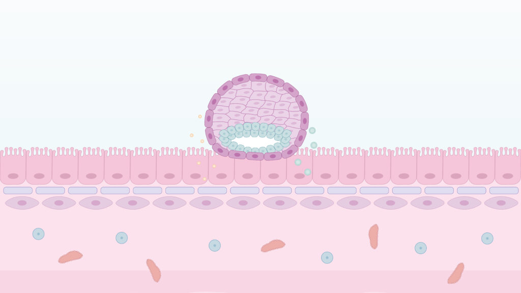
A few days after fertilization, the human embryo reaches the uterus at the blastocyst stage, around days 5 to 6 after fertilization. To establish pregnancy, the blastocyst must first establish contact with the endometrium, the tissue lining the inside of the uterus. The embryo approaches the endometrial surface in the correct orientation (apposition), then attaches to it (adhesion) and, later, begins to invade the underlying tissue (invasion).
Successful pregnancy depends on both the embryo and the endometrium. The embryo needs to be properly developed and capable of implantation, while the endometrium must reach a receptive state. Understanding how the embryo and endometrium interact during implantation is important, as alterations in this process can contribute to implantation failure.
Yet studying human implantation remains challenging, as it takes place inside the uterus and cannot be directly observe the process directly as it unfolds.
To address this challenge, our team has developed an in vitro model of the human endometrium to study the adhesion stage of implantation. The work, published in Science Advances, showed that the model recapitulates key features of human embryo adhesion.
Implantation begins before the embryo attaches
The endometrium is a highly dynamic tissue that undergoes continuous changes in response to steroid hormones. These changes prepare the endometrium to receive the embryo and support implantation.
During the secretory phase of the menstrual cycle, when the endometrium prepares for a potential pregnancy, endometrial epithelial cells acquire characteristics associated with receptivity, creating a favorable environment for embryo attachment.
At the same time, stromal cells beneath the epithelium undergo a process known as decidualization. During this process, the cells change their structure and function to help create an environment capable of supporting implantation and early pregnancy.
These changes involve coordinated communication between different cell types within the endometrium. Together, they create the cellular and molecular environment required for the embryo to attach.
Recreating the endometrium on a chip
To model the adhesion phase of human implantation, the team developed a microfluidic model called ADOC, short for Adhesion-Dynamics-On-a-Chip.
The model uses a commercially available empty Organ-Chip device from Emulate that contains two small channels separated by a porous membrane. ADOC contains in the upper channel endometrial epithelial cells derived from organoids, three-dimensional cell cultures that reproduce some features of the original tissue. And in the lower channel contains stromal cells, which form part of the supporting tissue of the endometrium.
The porous membrane allows molecules to move between the two compartments, enabling the cells to communicate while keeping each cell population physically separated.
The model was then exposed to hormonal stimulation designed to reproduce key features of the secretory phase, when the endometrium acquires characteristics associated with receptivity.
The cells responded accordingly. The epithelial compartment acquired features associated with receptivity, while the stromal cells showed signs of decidualization.
The team also analyzed gene activity in more than 28,000 individual cells using single-cell RNA sequencing. This technique allowed them to examine how different cell populations responded to hormonal stimulation. The results showed that their molecular profiles shifted toward states similar to those observed in the human endometrium during the secretory phase.
The model also reproduced another relevant feature of endometrial physiology: the release of extracellular vesicles. These small membrane-bound structures can carry molecules from one cell to another and have previously been associated with maternal-embryonic communication.
In ADOC, endometrial cells released extracellular vesicles containing microRNAs, small RNA molecules involved in regulating gene activity, that had previously been associated with the secretory phase of the endometrium.
Together, these features were important because the aim was not simply to provide a surface on which to place an embryo. The model needed to reproduce relevant biological characteristics of the tissue that the blastocyst encounters inside the uterus.
Studying human embryo adhesion in vitro
Once ADOC had been hormonally stimulated to reproduce features of the secretory phase, human blastocysts donated for research were introduced into the epithelial channel and monitored using time-lapse microscopy.
This made it possible to observe the process step by step.
As adhesion progressed, the blastocysts lost their characteristic spherical shape and adopted a more compact structure. Contact with the endometrium began at the side of the embryo where the inner cell mass is located. This region of the outer embryonic layer, known as the polar trophectoderm, later contributes to the formation of the placenta.
As attachment progressed, some cells in this outer layer became multinucleated, showing early features of the cellular changes associated with placental development. Meanwhile, the inner cell mass, the group of cells that will give rise to the embryo itself, reorganized into a radial structure.
The attached embryos also secreted beta-hCG, a hormone produced during the early stages of implantation.
Under hormonally prepared conditions, 9 of the 19 human blastocysts studied established stable adhesion. On average, adhesion began approximately 34 hours after the blastocysts were introduced into the model.
The hormonal environment proved essential. When blastocysts were introduced into models that had not received hormonal preparation, none established stable adhesion. Blocking the action of progesterone with mifepristone also markedly reduced adhesion, further supporting the importance of hormonal preparation of the endometrium for embryo attachment.
What can this model tell us about implantation?
ADOC does not reproduce the entire process of human implantation. The model focuses on embryo adhesion and does not capture the subsequent invasion of endometrial tissue or the full cellular and structural complexity of the human endometrium.
Instead, ADOC provides a controlled experimental system to study some of the key events that take place during the earliest stages of implantation. By recreating relevant features of the endometrium and combining them with human embryos, the model provides a way to examine a process that is normally hidden from direct observation inside the uterus.
This makes it possible to study in greater detail how the endometrium prepares for implantation, which changes accompany embryo adhesion, and which factors may affect this process.
Human implantation remains one of the least accessible stages of early pregnancy, and many aspects of the process are still not fully understood. Models such as ADOC provide a way to study human embryo adhesion under controlled conditions and better characterize the cellular and molecular events that accompany these early stages.
Article reference
Zaragozano S, Pardo-Figuerez M, Monteagudo-Sanchez A, et al. Modeling human embryo adhesion using a microfluidic platform. Science Advances. 2026;12:eadz2249. https://doi.org/10.1126/sciadv.adz2249