Assisted reproduction has transformed reproductive medicine since the birth of the first baby conceived through in vitro fertilization in 1978. Today, more than 10 million children worldwide are estimated to have been born thanks to IVF and other assisted reproductive technologies [1]. However, outcomes still depend on multiple factors, including age, the cause of infertility, and embryo quality.
One of the major challenges remains assessing which embryo has the greatest developmental potential before it is transferred to the uterus. This decision must be made as accurately as possible, while meeting one essential condition: obtaining useful information without harming the embryo.
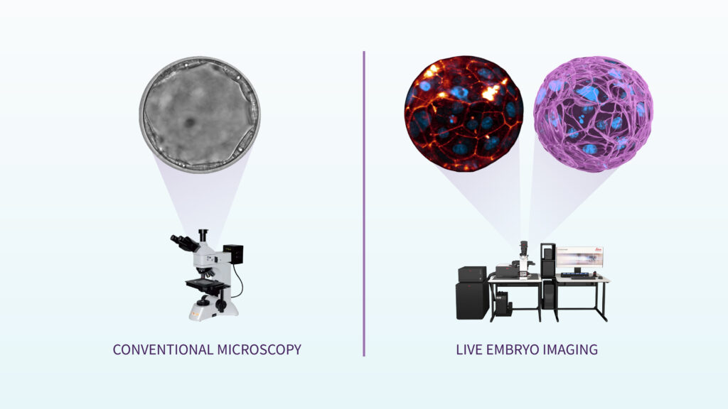
For years, embryo assessment has relied largely on morphological evaluation using conventional microscopy. This approach has been valuable in the IVF laboratory, but it provides a limited view of a process that unfolds in three dimensions and changes continuously over time.
An embryo is not a still image. During its first days of development, its cells divide, compact, change position, establish contacts with one another, and begin to differentiate into the first cellular structures. The inner cell mass will give rise to the embryo proper, while the trophectoderm will contribute to placenta formation. Recent live-imaging studies have made it possible to observe these processes in human embryos, including compaction, polarization, blastocyst formation, and hatching, the process by which the embryo leaves the zona pellucida before implantation [2].
New live-imaging technologies allow researchers to study these processes with higher resolution and in real time. By combining advanced microscopy, fluorescent markers, and computational analysis in experimental settings, researchers can observe how the embryo organizes before implantation and identify cellular behaviors that may go unnoticed during conventional evaluation. These include errors in cell division, alterations in chromosome segregation, micronuclei, binucleated cells, and other behaviors associated with defects in early embryonic development [3].
This is the foundation of the Carlos Simon Foundation’s embryo imaging research line, which focuses on improving embryo quality assessment by studying preimplantation development in mouse embryos and, subsequently, in human embryos donated for research.
The goal is not only to see better, but to understand better. Observing how cells divide, how genetic material is organized, how embryo integrity is maintained, and how the blastocyst forms may help identify signals associated with normal or altered development.
In the future, this knowledge could contribute to the development of more precise, non-invasive methods to assess embryo viability before transfer. However, its clinical application requires caution: these technologies still need to be thoroughly validated before they can be incorporated into routine practice.
References:
- European Society of Human Reproduction and Embryology. ART Fact Sheet. ESHRE; 2025. https://www.eshre.eu/-/media/sitecore-files/Press-room/ESHRE_ARTFactSheet_v10_2025.pdf
- Domingo-Muelas A, et al. Human embryo live-imaging reveals nuclear DNA shedding during blastocyst expansion and biopsy. Cell. 2023. https://doi.org/10.1016/j.cell.2023.06.003
- Akizawa H, et al. Capturing aberrant cell behaviors producing defects in human embryos via live imaging. Science Advances. 2025. https://doi.org/10.1126/sciadv.ady6402